Biology (SJCR)
Stechnolock Journal of Case Reports
Full Text
Volume 1, Issue 1
Synchronous Bilateral Ovarians Tumors: Mixed Müllerian Ovary Tumor Associated with Serious Adenocarcinoma of Controlateral Ovarius: A Case Report
*Corresponding Author: Moukhlis E, Gynecological Service, IBN ROCHD University Hospital Center, Faculty of Medicine and Pharmacy, Hassan 2 University Casablanca, Morocco, Tel: +212600882164, E-mail: sabah.moukhlis@gmail.com
doi: /sjcr.2021.1.105
Citation: Oubid A, Moukhlis S, Elhachami F, Boufettal H, Mahdaoui S, et al. (2021) Synchronous Bilateral Ovarians Tumors: Mixed Müllerian Ovary Tumor Associated with Serious Adenocarcinoma of Controlateral Ovarius: A Case Report. Stechnolock J Case Rep 1: 1-5
Copyright: © 2021 Oubid A, Moukhlis S. This is an open-access article distributed under the terms of Creative Commons Attribution License, which permits unrestricted use, distribution, and reproduction in any medium, provided the original author and source are credited.
.JPG)
.JPG)
Abstract
Mixed Mullerian tumor also called ovarian carcinosarcoma or mixed mesodermal tumor is among the rare malignant tumors affecting the ovary which associates a carcinomatous component with a sarcomatous component. It is a rare tumor known to be highly aggressive and typically affects postmenopausal women between the ages of 60 and 70. And if this type of tumor is rare, its synchronous development with a contralateral ovarian tumor is exceptional.
In this work, we report a case of a patient who presented with abdominal distension and whose positive diagnosis was obtained postoperatively on the operative specimen of a right ovarian carcinosarcoma associated with an invasive papillary serous adenocarcinoma of the left ovary. Through this observation and a brief review of the literature, we specify the epidemiological, anatomopathological, clinical, radiological characteristics and the therapeutic management of synchronous ovarian tumors.
Keywords: Ovarian Carcinosarcoma; Synchronous Ovarian Tumors; Serous Adenocarcinoma of The Ovary
Introduction
Carcinosarcoma is an aggressive tumor that combines a carcinomatous component with a sarcomatous component. It is most often found in the body of the uterus. The ovary, cervix and vagina are more rarely affected [1]. The only prognostic factor found is the initial stage of the tumor.
Ovarian carcinosarcoma (OCC) affects women, most often nulliparous between the sixth and seventh decades. Age at diagnosis is significantly higher in ovarian carcinosarcoma than in ovarian epithelial tumor [2].
The coexistence of cancers in two histologically different ovaries remains rare. Through this observation and a brief review of the literature, we specify the epidemiological, anatomopathological, clinical, radiological characteristics and the therapeutic management of synchronous ovarian tumors.
Case presentation
A woman, 65 years old, married, menopausal for 15 years without a particular pathological history, had for six months an abdominal distension with chronic pelvic pain without other associated signs, particularly digestive or urinary, the clinical examination found on palpation an abdominal mass. Pelvic arriving at the umbilicus and vaginal touch the perception of the lower pole of the mass.
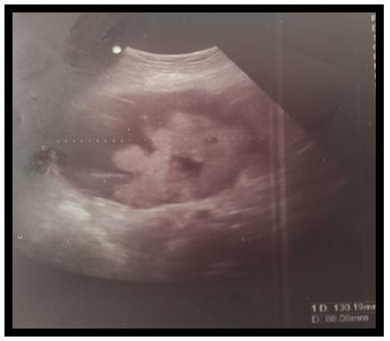
The pelvic ultrasound showed in the right latero uterine a heterogeneous echostructural mass with a large fleshy part, measuring 130 mm x86 mm reaching the umbilicus with evidence in the left sub pelvic of a cystic mass with septum and fleshy component 85 x 67 mm (Figure 1).
The abdominal pelvic CT scan revealed a moderate peritoneal effusion with two bilateral ovarian masses with abdomino-pelvic development with double cystic and solid contingent, well limited by regular contours, the most voluminous on the right measuring approximately 14x12x17 cm and of which the cystic component is hypodense seat of thin partitions within it which are enhanced after injection of contrast product (Figure 2).
A digestive exploration made by an eso-gastro-duodenal fibroscopy was performed showed a sliding hiatus hernia and atrophic antral gastritis without other abnormalities.
Tumor markers were 375.7 UI / ml positive for CA125 and CA19-9 was 116.2 U / ml.
Surgical exploration revealed two solido-cystic latero-uterine masses on the right of 10cm and on the left of 12cm. Bilateral adnexectomy was performed and sent for extemporaneous examination:
Bilateral borderline serous tumor with foci of invasion. A total hysterectomy was subsequently performed with omentectomy and bilateral lymph node pelvic dissection.
The final histological examination of the specimens found in the right adnex a mixed malignant mullerian tumor with a predominantly serous-type epithelial component measuring 15cm, and in the left adnex a high-grade invasive papillary serous adenocarcinoma measuring 14cm.
Discussion
The diagnosis of ovarian cancers presents one of the most difficult problems because they are silent, their symptoms are varied and nonspecific, and clinical signs appear at an advanced stage. The clinical presentation of OCS ovarian carcinosarcoma is nonspecific. The most common symptom is abdominal distension. It may be associated with abdominal pain, transit disorders and a deterioration in general condition. Very often the diagnosis is made at an advanced stage of the disease. Neither do metastatic locations differ from those of TEO epithelial tumors of the ovary [2].
Coexistence of ovarian and endometrial tumors is common, but the synchronous development of ovarian cancers is rare, therefore, data available in the literature for the management of these cases are limited. About ten case reports have been published which presented cases of Mullerian ovarian tumors associated with tumors in the endometrium or other sites in the female genital tract, but only one case similar to ours has been described previously from first diagnosis until death [3,4]: a 58-year-old woman with bilateral synchronous ovarian malignancies.
Rewsuwan S et al published in 2016 a case of ovarian carcinosarcoma associated with mature cystic teratoma and primary tubal carcinoma [5]. Arora et al. reported a case of Mullerian tumor of the broad ligament in association with uterine endometrioid adenocarcinoma associated with serous papillary ovarian carcinoma [6].
The initial stage is the only prognostic factor found in the various studies. The more advanced the stage, the worse the prognosis. At the same stage, OCS has a worse prognosis than epithelial tumors of the ovary. Brown is the only one to have compared these two tumors. He found a significant difference in the median survival (8.2 versus 20.7 months, p <0.0001), progression-free survival (6.4 versus 12 months, one month, p = 0.001), and survival at five years in favor of TEOs [2].
The rarity of the CSO explains why there is no consensus on its management. There is very little data [2]. For adjuvant therapy, the only published trial is that of Tate Thigpen for the Gynecologic Oncology Group. The substance used is cisplatin. The response rate is 20%, which is comparable to that observed in uterine CS [7].
The surgical exploration has three objectives: Histological diagnosis, Surgical and therapeutic staging (tumor reduction).
Aggressive cytoreductive surgery appears to have a positive impact on the outcome and should probably be offered to most patients [8].
Conclusion
Primary malignant ovarian tumors of a different histological nature in each ovary is a particularly interesting situation because the information available about it is scarce and because there is little consensus on its definition, classification and correct diagnostic approach- therapeutic.
- Regional Cancer Network of Lower Normandy "OncoBN" (2017) Repository rare malignant tumors of the ovary diagnostic and therapeutic management, Morocco.
- Muller M (2007) ovarian carcinosarcoma. Journal of Obstetrics Gynecology and Biology of Reproduction 36: 399-402.
- Günakan E, Tohma YA, Haberal AN, Ayhan A (2018) Bilateral synchronous ovarian tumours: an uncommon case and review of the literature. Menopause Rev 17: 97-100.
- Chanson MJ, Lee CW, Seo KJ, Kim JA, Parc JS, et al. (2011) A case of bilateral ovarian synchronous tumors (left ovarian serous papillary adenocarcinoma and right ovarian malignant mixed Müllerian tumor [Un cas de tumeurs synchrones ovariennes bilatérales (adénocarcinome papillaire séreux ovarien gauche et tumeur Müllérienne mixte maligne ovarienne droite)]. Eur J Gynecol Oncol 32: 234-6.
- Rewsuwan S, Satabongkoch N (2016) Ovarian Carcinosarcoma and Its Association with Mature Cystic Teratoma and Primary Tubal Carcinoma 2016: 2605045.
- Arora P, Rao S, Khurana N, Talwar D (2011) Malignant mixed Mullerian tumor of broad ligament with synchronous ovarian and endometrial carcinoma: A rare association. J Canc Res Therap 7.
- Berroho K, Achiq A, Fillali A (2011) Ovarian carcinosarcoma: one case report [Carcinosarcome ovarien : à propos d’un cas]. J Afr Cancer 3: 147-50.
- Mano MS, Rosa DD, Azambuja E (2007) Current management of ovarian carcinosarcoma. Int J Gynecol Cancer 17: 316-24.